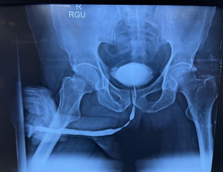
Retrograde Urethrography Surgery in Pune
Blockages and injuries to the urethra can be serious and often require immediate medical attention. One of the key diagnostic tools used to evaluate such issues is retrograde urethrography (RUG).
This article will delve into what a retrograde urethrography, why is it performed, the procedure itself, and what patients can expect.
Additionally, we will address some frequently asked questions to provide a well-rounded understanding of this important diagnostic tool.
What is a Retrograde Urethrogram?
A retrograde urethrogram is a specialized imaging technique used to visualize the urethra and assess for any injuries or abnormalities.
It involves the injection of a contrast dye into the urethra, allowing healthcare providers to capture detailed X-ray images of the urethra’s structure.This procedure is particularly important in cases of trauma, where injuries may not be immediately apparent.
Why is a Retrograde Urethrogram Performed?
Traumatic retrograde urethrograms are primarily indicated in situations where there is a suspicion of urethral injury, particularly following pelvic fractures or other significant trauma. Common reasons for performing a RUG include:
– Pelvic Trauma: Injuries from car accidents, falls, or sports-related incidents can lead to urethral damage.
– Blood at the Urethral Meatus: The presence of blood at the opening of the urethra is a strong indicator that a urethral injury may have occurred, warranting further investigation.
– Difficulty Urinating: Patients experiencing urinary retention or difficulty voiding may require a RUG to determine if an obstruction or injury is present.
– Post-Surgical Evaluation: After certain surgical procedures, a RUG may be performed to assess the integrity of the urethra.
The Procedure: What to Expect
Preparation
Before undergoing a traumatic retrograde urethrogram, patients will typically receive specific instructions. These may include:
– Fasting: Patients may be asked to refrain from eating or drinking for a few hours before the procedure.
– Medication Review: It’s essential to inform the healthcare provider of any medications being taken, especially blood thinners.
– Urine Sample: A urine sample may be collected to check for infections that could complicate the procedure.

During the Procedure
The retrograde urethrogram is usually performed in a radiology department or emergency setting. Here’s what patients can expect during the procedure:
- Positioning: Patients will lie on an examination table, typically in a supine position (on their back).
- Anesthesia: A local anesthetic gel may be applied to the urethra to minimize discomfort during catheter insertion.
- Catheter Insertion: A thin catheter is gently inserted into the urethra. This catheter is connected to a syringe filled with contrast dye.
- Contrast Injection: The healthcare provider slowly injects the contrast dye into the urethra. As the dye fills the urethra, X-ray images are taken to visualize its structure.
- Image Capture: The radiologist will capture a series of images as the contrast material fills the urethra. Patients may be asked to hold their breath briefly during this process.
After the Procedure
Once the procedure is complete, patients will be monitored for a short time. Some mild discomfort, such as a burning sensation during urination, is common but usually temporary. Drinking plenty of fluids can help flush out the contrast dye and alleviate discomfort.
Understanding the Results
The results of a traumatic retrograde urethrogram can provide critical information regarding the presence and extent of urethral injuries:
– Normal Results: Indicate that there are no visible injuries or abnormalities in the urethra.
– Abnormal Results: May reveal conditions such as:
– Urethral Strictures: Narrowing of the urethra that can impede urine flow.
– Urethral Rupture: A complete or partial tear in the urethra, often requiring surgical intervention.
– Extravasation: Leakage of urine or contrast dye into surrounding tissues, indicating a serious injury.
Healthcare providers will discuss the results with patients, explaining what they mean in the context of their symptoms and overall health.
Potential Risks and Complications
While a retrograde urethrogram is generally safe, there are some risks associated with the procedure, including:
– Infection: There is a small risk of urinary tract infection following the procedure.
– Bleeding: Some patients may experience minor bleeding from the urethra.
– Discomfort: Temporary discomfort or burning sensation during urination is common.
Patients should contact their healthcare provider if they experience severe pain, significant bleeding, or signs of infection (such as fever or chills) after the procedure.
FAQs
Most patients experience only mild discomfort during the procedure, especially during catheter insertion. A local anesthetic is used to minimize pain
A retrograde urethrogram typically takes about 30 minutes to an hour, including preparation and imaging time.
Patients may be instructed to avoid eating or drinking for a few hours before the procedure. It’s essential to follow your healthcare provider’s specific instructions.
Your healthcare provider will usually discuss the results with you shortly after the procedure, although it may take a few days for the final report to be available.
Other imaging techniques, such as ultrasound or MRI, may be used to evaluate the urinary tract, but a retrograde urethrogram is often preferred for detailed visualization of the urethra.
Conclusion
A retrograde urethrogram is a vital diagnostic tool in the assessment of urethral injuries. Understanding the procedure, its purpose, and what to expect can help alleviate any concerns.
If you suspect a urethral injury or are experiencing urinary issues, consult your healthcare provider for a thorough evaluation and appropriate treatment options.Taking proactive steps in understanding your health is always a positive move!
